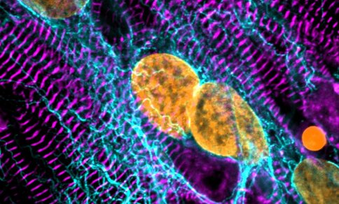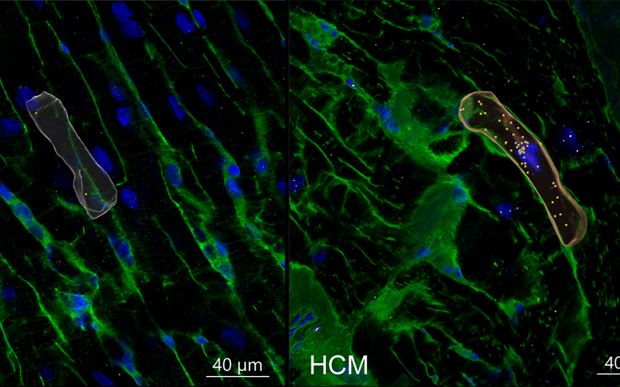
Heart muscle disease comes in many forms, the most common of which are hypertrophic cardiomyopathy (HCM), which causes the heart muscle to become thick and stiff, and dilated cardiomyopathy (DCM), in which the left ventricle balloons, thinning the chamber’s walls. Both types make it hard for the heart to pump properly and can eventually cause it to fail.
A team led by researchers at the Max Delbrück Center and Brigham and Women’s Hospital now shows in “Science Translational Medicine” the detailed molecular activity underlying HCM, including which pathways it shares with DCM.What’s more, they were able to distinguish early stage from late stage, and genetic from non-genetic HCM.
The research was co-led by Dr. Eleonora Adami (Max Delbrück Center, Hübner lab) and Dr. Yuri Kim (Brigham and Women’s Hospital) as part of an international collaboration that included researchers at Harvard (Seidman Lab), Imperial College London (Noseda Lab), the University of Alberta (Oudit Lab), Helmholtz Munich, TU Munich (Heinig Lab), and Herz und Diabeteszentrum NRW (Milting Lab).
Studying disease one cell at a time
To characterize the cellular and molecular signatures of HCM, the researchers used single-nucleus RNA sequencing to analyze heart tissue from 47 patients spanning early stage to end-stage HCM, including people with non-genetic forms of the disease. The researchers then compared these gene expression profiles with data from healthy donor hearts and heart tissue affected by DCM.
The technology allowed them to document which genes were active in nearly one million individual heart cells. They created a comprehensive molecular map detailing gene activity according to stage of disease and genetic status.
They found a few “surprising” differences, says Adami. In early-stage HCM, for example, fibroblasts — which produce and maintain the extracellular matrix that surrounds cells — expressed less collagen IV. In effect, this would likely destabilize the matrix. They also found that patients with genetic HCM had proportionally fewer cardiomyocytes compared to healthy donors, along with elevated expression of genes related to irregular heartbeat and scar tissue formation.
“It has long been known that HCM patients with a known genetic cause tend to suffer more severe disease, yet the molecular mechanisms accounting for this have remained unknown,” says Adami. “We show that these patients have a distinct pattern of gene activity in their hearts.”
Additionally, the researchers identified the gene PRR16 as a potential contributor to the enlarged muscle cells that are characteristic of HCM.
HCM, not just a disease of muscle cells
In a final step, the team showed that an AI model trained on the gene expression data from heart muscle cells could accurately distinguish early stage from late-stage HCM, separate HCM from DCM, and correctly identify patients with genetic versus non-genetic HCM.
Based on gene expression data from fibroblasts alone, the model was also able to make the same distinctions. “This was interesting,” explains Kim, “because cardiomyopathies are usually thought of as diseases of heart muscle cells only.”
“It was intriguing to see that the unbiased multiomics factor analysis revealed that so many different cell types contribute to the signatures that distinguish between patient groups and controls,” says Dr. Matthias Heinig, an author at Helmholtz Munich and TU Munich.
“By mapping gene expression at single-cell resolution across disease stages and genetic subtypes, we’ve built a molecular signature of HCM’s clinical spectrum,” Hübner adds. “This should provide a foundation for future work on more targeted treatments.”
Source: Max Delbrück Center press release
Original publication: Adami E, Kim Y, Zheng SL, et al. The molecular landscape of hypertrophic cardiomyopathy across disease stages and genotypes. Sci Transl Med. 2026;18(867):eaea2747. doi:10.1126/scitranslmed.aea2747